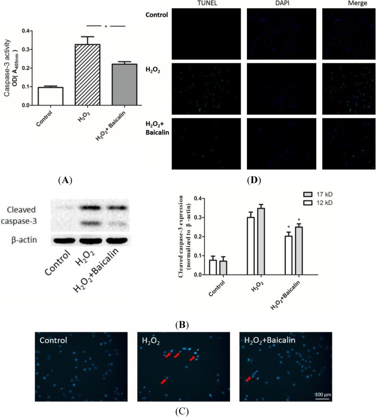
Figure 3.
Effects of Baicalin on H2O2-induced apoptosis in HK-2 cells. The activity of caspase-3 (A) and the expression of cleaved caspase-3 (B) were evaluated in cultured HK-2 cells. H2O2 treatment up-regulated both the activity of caspase-3 and the expression of cleaved caspase-3. Baicalin pretreatment suppressed H2O2 and induced up-regulation of caspase-3 activity and cleavage; Cultured HK-2 cells were stained with Hoechst (C) Apoptotic cells showed nucleic fragmentation with dense chromatin (red arrow); The transferase-mediated dUTP-biotin nick end labeling (TUNEL) assay revealed more positive staining cells (with green fluorescence) in the H2O2 group than in the H2O2 + Baicalin group (D). H2O2 treatment increased cell apoptosis, while Baicalin pretreatment suppressed cell apoptosis. * p < 0.05, vs. the group with H2O2 treatment, n = 6.

