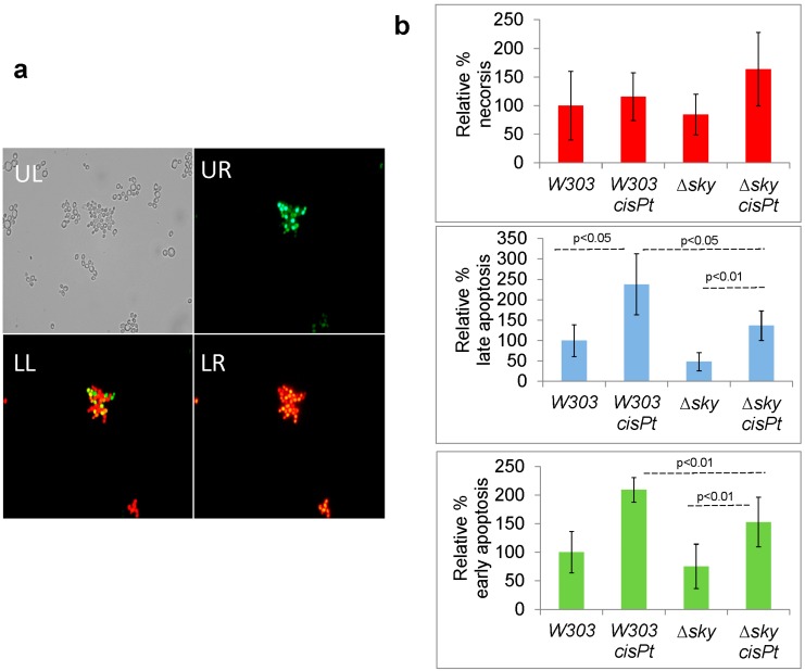
Figure 3.
Apoptosis and necrosis induced by cisplatin (cisPt) in W303-1A (W303) and ∆sky1 strains (a) apoptosis or/and necrosis visualized by fluorescence microscopy (objective 40×): UL, Bright field; UR Green channel for YO-PRO®-1 staining; LR, Red channel for PI staining; LL Merge; (b) Quantification of necrosis, late apoptosis and early apoptosis by flow cytometry.

