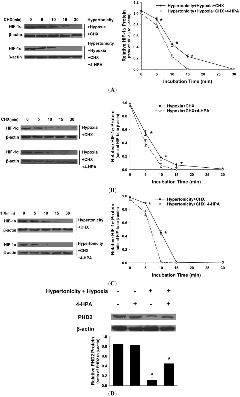
Figure 7.
Effects of 4-HPA on HIF-1α protein degradation under hypertonicty and hypoxia conditions in primary AEC. AEC were treated with hypertonicity (C); hypoxia (B); or both of the two (A) with or without 4-HPA treatment for 4 h, followed by incubation with 100 μM cycloheximide (CHX, blocking ongoing protein synthesis) from 0–30 min. Cell lysates were subjected to Western blotting using antibodies against HIF-1α and β-actin (the left panel) and the intensity of HIF-1α protein relative content was quantified (the right panel). The plot represented means ± SD from three independent experiments, * p < 0.05 vs. groups with 4-HPA treatment; and (D) prolyl hydroxylase domain enzyme isoform-2 (PHD2) protein of AEC which were treated with both hypertonicity and hypoxia in the presence and absence of 4-HPA was detected by Western blotting. Data are means ± SD, & p < 0.01 vs. control group, # p < 0.05 vs. hypertonicity + hypoxia group.

