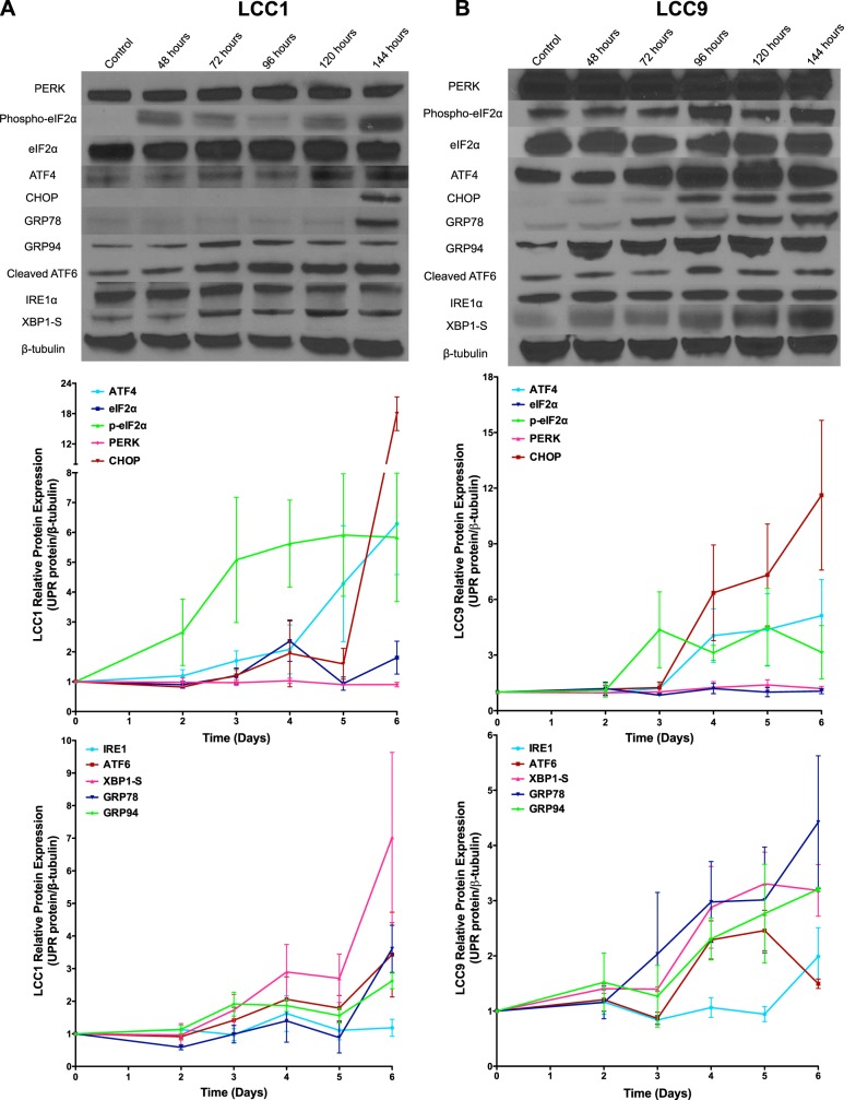
Figure 1.
ICI differentially activates UPR signaling in antiestrogen-sensitive LCC1 and antiestrogen-resistant LCC9 cells. Representative Western blot images of PERK, phospho-eIF2α, eIF2α, ATF4, CHOP, IRE1α, XBP1-S, cleaved-ATF6, GRP78, GRP94, and β-tubulin (loading control) in LCC1 (A) and LCC9 (B) breast cancer cells treated with 100 nM ICI for 6 d. Biological replicate Western blots (n = 3) for each time course were quantified with ImageJ, and data are reported as relative protein expression.

