Figure 5.
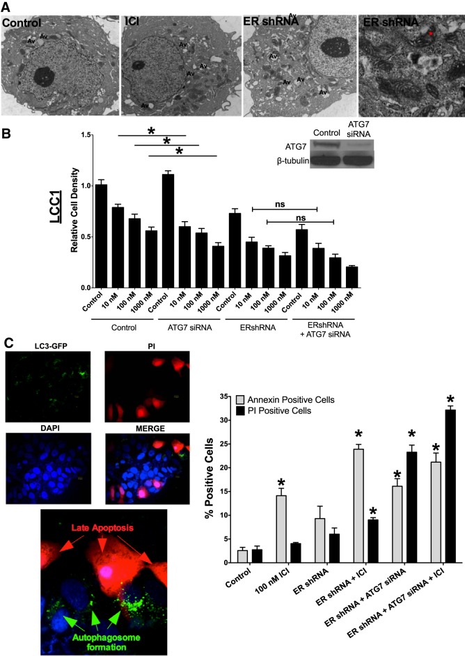
ERα-mediated autophagy is prosurvival. A) EM of LCC9 breast cancer cells indicated elevated autophagy and mitochondrial lipid deposition with ERα knockdown. LCC9 breast cancer cells were treated with vehicle, ICI, or ERshRNA and were used to determine cellular morphology by EM. Asterisk: lipid deposition in the mitochondria. Av, autophagic vacuoles. B) LCC1 cells were transfected with control (control siRNA + control shRNA) or ERα shRNA and/or ATG7 siRNA and treated with 0.1% v/v ethanol vehicle or 10, 100, and 1000 nM ICI for 72 h, and cell density was measured with crystal violet. Western blot hybridization of treated protein homogenates was used to measure ATG7 and β-tubulin. C) LCC1 cells were transfected with GFP-LC3 and ERα shRNA and treated with 100 nM ICI for 72 h. The cells were stained with PI and counterstained with DAPI. LC3 puncta and PI positivity were confirmed by confocal microscopy. D) LCC1 cells were transfected with control siRNA or ERα shRNA and/or ATG7 siRNA and treated with 100 nM ICI for 72 h. Annexin V-FITC and PI-stained cells were counted by using flow cytometry (n = 3). *P < 0.05; 1-way ANOVA with the Bonferroni post hoc analysis.

