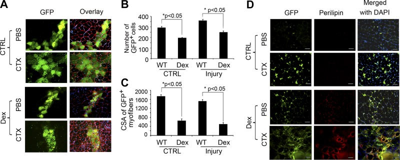
Figure 3.

In vivo, Dex stimulates FAPs to differentiate into adipocytes. A) At 10 d after the transplantation of satellite cells isolated from EGFP mice into muscle, GFP-expressing myofibers were found along the needle track; laminin identifies the basement membranes of myofibers. B) Numbers of green myofibers were counted. C) Sizes of green myofibers were measured (n=100 myofibers). D) FAPs isolated from EGFP mice were injected into TA muscles of mice that had been treated with Dex or CTX to induce muscle injury (bottom panels). Cross-sections were immunostained with anti-perilipin (red) and GFP. Scale bars = 50 μm.
