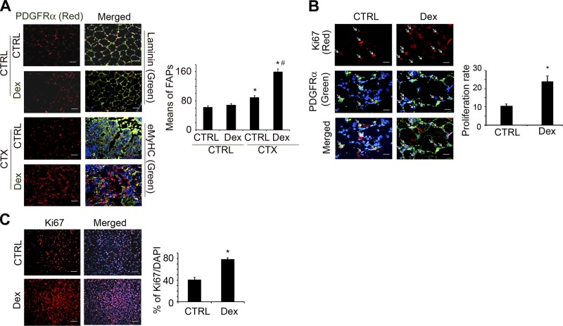
Figure 4.
Dex stimulates FAP proliferation in vivo and in vitro. A) FAPs were evaluated by immunostaining muscle cross-sections with anti- PDGFRα and anti-laminin (uninjured muscles) or with anti-eMyHC (injured muscles). In sections from Dex-treated and control mice, means of PDGFRα cells per view were counted (right panel). *P < 0.05 vs. no injury/no Dex; #P < 0.05 vs. Dex no injury. B) FAP proliferation at 3 d after muscle injury was evaluated by coimmunostaining of cross-sections of muscles with anti-Ki67 and anti-PDGFRα. Right panel shows percentage of double-positive cells vs. PDGFRα+ cells. *P < 0.05 vs. control (Ctrl), no Dex. C) Cultured FAPs were stimulated with Dex (1 μM) for 24 h and then immunostained with anti-Ki67. Right panel shows proliferation rates as percentage of Ki67+ cells to total number of cells (n=3 repeats). *P < 0.05 vs. control, no Dex.

