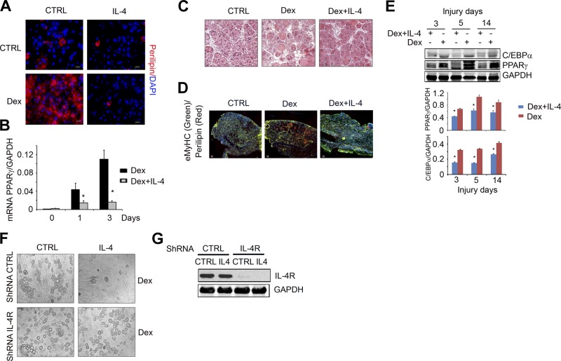
Figure 6.
IL-4 suppresses Dex-induced FAP adipogenic differentiation. A) FAPs were isolated by FACS and placed in GM for 7 d and then cultured in medium with Dex (1 μM) or Dex plus IL-4 for 4 d. Control (Ctrl) cells were continually cultured in GM. The fixed cells were immunostained with anti-perilipin (red) and DAPI (blue). B) In FAPs using similar treatment as in panel A, mRNA of PPARγ was evaluated by RT-PCR (n=3 repeat). *P < 0.05 vs. Ctrl. C) C57/BL6 mice were treated with Dex for 4 d and followed treatment with Dex plus IL-4 for 1 d before TA muscles were injured by CTX injection. At 8 d after muscle injury, cryo-cross-sections of TA muscles were H&E stained. D) Cross-sections of injured TA muscle from panel C were coimmunostained with anti-perilipin (red) plus anti-eMyHC (green). Scale bars = 50 μm. E) Top panel shows representative Western blots show the levels of C/EBPα and PPARγ in injured muscle of mice treated with Dex or Dex plus IL-4 at different days after injury. Bottom panel shows density (n=4). *P < 0.05 vs. Dex alone. F) FAPs were transfected with lentivirus of shRNA of IL-4R, or shRNA CTRL. After 4 d, cells were treated with Dex adipogenic differentiation medium with or without IL-4 for 7 d, and cells were photographed under light microscopy. G) Representative Western blots show levels of IL-4R in the cells from panel F.

