
Figure 2.
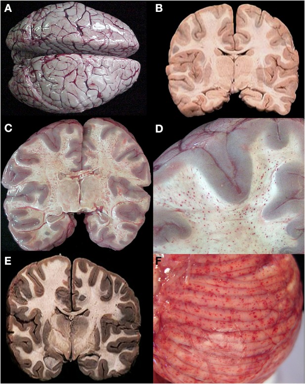
A summary of representative images from the gross examination of the brain in the autopsy series is shown for CM cases. The vast majority of cases, regardless of diagnosis, showed brain swelling with flattened gyri and narrowed sulci (A). In this example, the brain has the classic “slate gray” to “purple” appearance of CM which is possibly due to malaria pigment within vessels. In all control cases and in a subset of the CM1 cases, the coronal brain slice appeared without discoloration (B). In the classic CM2 appearance, petechial hemorrhages are seen diffusely in the white matter throughout the brain (C). A higher magnification demonstrates the abrupt transition from white to gray matter and the lack of hemorrhages in the gray (D). In a subset of the CM1 cases, the coronal brain slice appeared very discolored (an unexplained phenmenon) and swollen but without petechial hemorrhages in the cerebral cortex (E). The cerebellum had petechial hemorrhages in both the gray and white matter and thus, visible on the surface grossly (F). (B,E) Were previously published in part in the Journal of Infectious Diseases (2005) and appear here with express permission (see Milner et al., 2005; White and Silamut, 2005).