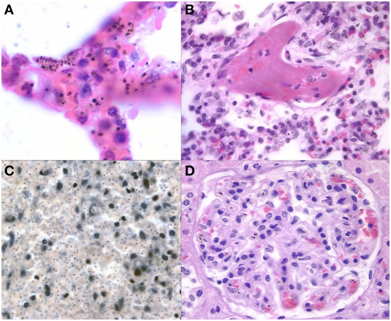
Figure 7.
Representative images of pathology outside of the brain are shown. The lungs (A) demonstrate some sequestration of parasites but more frequently have heavily burdened macrophages with variable size pigment globules (1000X, H&E). Fibrin thrombi (B) were present in a subset of CM2 and other cases in the lungs suggestive of DIC (400X, H&E). The spleen (C) in a patient with CM after depigmentation of the section using picric acid followed by Giemsa stain (from Milner, unpublished data) demonstrating the unique finding of dense parasite burdens in CM1 spleens (200X, Giemsa). A representative glomerulus (D) from the kidney shows malaria pigment within macrophages and microthrombi present in capillaries (400X, H&E).

