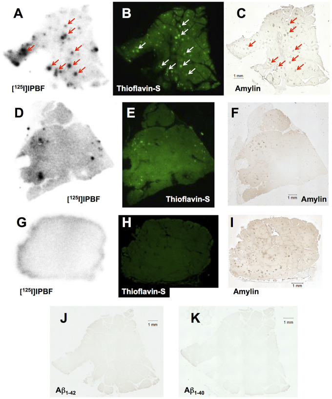
Figure 5. Comparison of autoradiographic images of [125I]IPBF with pancreatic tissue sections from a T2DM patient and two healthy controls (A, and D and G, respectively).
The same sections were also stained with thioflavin-S (B, and E and H, respectively). Immunohistochemical staining with antibodies against amylin (C, and F and I, respectively), Aβ1-42 (J), and Aβ1-40 (K). Pancreatic tissue sections from a T2DM patient (A, B, C, J, and K) and two different healthy donors (D, E, and F: 71-year-old man; G, H, and I: 28-year-old man).

