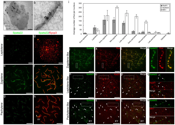
Figure 1. Spata22 colocalises with Rpa in meiotic nodules.
(a, b) Immunoelectron microscopy of a pachytene spermatocyte nucleus. Higher magnification of the SC indicated by an arrow in (a) (b). Spata22 localised to meiotic nodules (c–h). Distribution of Spata22 in spermatocyte nuclei during leptotene (c, d), zygotene (e, f), and pachytene (g, h). Punctate staining signals of Spata22 (green) were distributed along axial/lateral elements (red). Strong immunofluorescent signals were present in the terminal segments of the sex chromosomes (XY) in the pachytene nucleus ((g), (h), arrows). (i) Average number of Spata22, Rad51, and Mlh1 foci in each substage of meiotic prophase I. Bars, standard deviations. (j–u) Comparison of the distribution of Spata22 foci (green) with the distribution of Rpa, Rad51, and Mlh1 foci (red) in the spermatocyte nuclei. Overlap of the two immunofluorescent signals is indicated in yellow. Spata22 foci and Rpa foci coexisted almost completely in the zygotene-like nucleus (j–m). Spata22 only, Rpa only, and the overlap were 0.8%, 1.1%, and 98.1% of all the foci in the nucleus, respectively. Higher magnification of the white boxes in (j–l) shows that Spata22 foci overlapped almost completely with Rpa foci (m). Rad51 foci and Spata22 foci partially coexisted in the leptotene-like nucleus (n–q). Spata22 only, Rad51 only, and the overlap were 18.2%, 60.1%, and 21.7% of all the foci in the nucleus, respectively. Complete overlap, partial overlap, and non-overlap of the two proteins' foci are indicated by large arrowheads, small arrowheads, and arrows, respectively. Higher magnification of the white boxes in (n–p) shows that Rad51 foci partially overlapped with Spata22 foci ((q), arrowheads). Mlh1 foci and Spata22 foci partially coexisted in the pachytene-like nucleus (r–u). Spata22 only, Mlh1 only, and the overlap were 83.3%, 4.4%, and 12.3% of all the foci in the nucleus, respectively. Complete or partial overlap and non-overlap between the two proteins' foci are indicated by arrowheads and arrows, respectively (r–t). Higher magnification of the white boxes in (r–t) shows that Mlh1 foci overlapped partially with Spata22 foci ((u), arrowheads). Scale bars in all panels, 10 μm.

