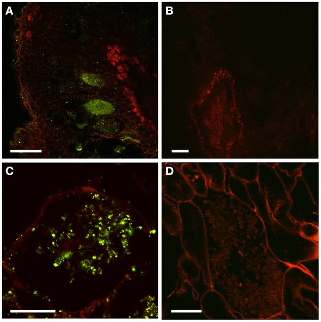
Figure 2.

Immunolocalization of PAA in D. trinervis nodules. (A–C). A strong signal is detected in cells infected by Frankia. No signal is present in the vascular bundle or in non-infected cells. (B–D) No signal is detected in control sections incubated with the secondary antibody alone. Scale bars: A, B: = 50 μm; C, D: 25 μm.
