Figure 5.
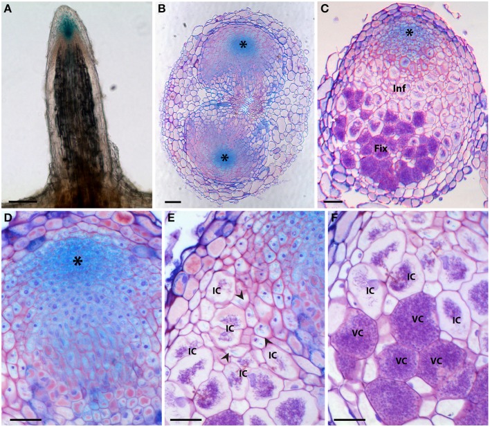
Histochemical localization of β-glucuronidase (GUS) activity in D. trinervis roots expressing a ProDtAUX1:GUS construct. (A) Non-inoculated lateral root; blue staining is detected in the root tip. (B) Cross section of an inoculated root 5 days after inoculation (dai) showing two nodule primordia growing from the pericycle at opposite xylem poles. DtAUX1 expression is observed in the meristematic cells (asterisk). (C) Longitudinal section of a fully developed nodule 21 dai. Cells containing Frankia hyphae are stained in purple. GUS activity is intense in the meristematic region, still visible in the infection zone (Inf), in non-infected cells, and non-detectable in the fixation zone (Fix). (D–E) Magnified images of (C). (D) Meristematic zone. (E) Infection zone: DtAUX1 expression is limited to non-infected cortical cells (arrowheads) surrounding the highly hypertrophied infected cells (IC). (F) Fixation zone: no DtAUX1 activation is detected in the hypertrophic cells filled with Frankia that have already differentiated nitrogen-fixing vesicles (VC). Sections (B–F) were stained with toluidine blue. Scale bars: 100 μm (A–C), 50 μm (D–F).

