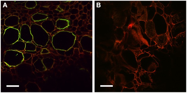
Figure 6.

Immunolocalization of PIN1-like proteins. (A) Longitudinal section of a mature nodule incubated with Anti-PIN1 antibodies and FITC labeled secondary antibodies: a strong signal is detected in the plasma membrane of hypertrophied cortical cells infected by Frankia. (B) Control section incubated with the secondary antibody alone where no signal is detectable. Scale bars: 50 μm.
