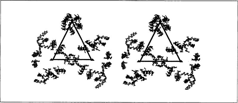
Fig. 2.
Stereoview of the γ-peptides and ordered RNA segments of the FHV particle structure. The triangle represents the border of the icosahedral asymmetric unit containing A, B and C in the T= 3 particle in Fig. 1. Blue helices are associated with the A subunits (γA), red with the B subunits (γB), and green with the C subunits (γc). Quasi six-fold axes are shown as red triangles, the five-fold axes as a red pentagon. Duplex RNA is represented as ball-and-stick models.

