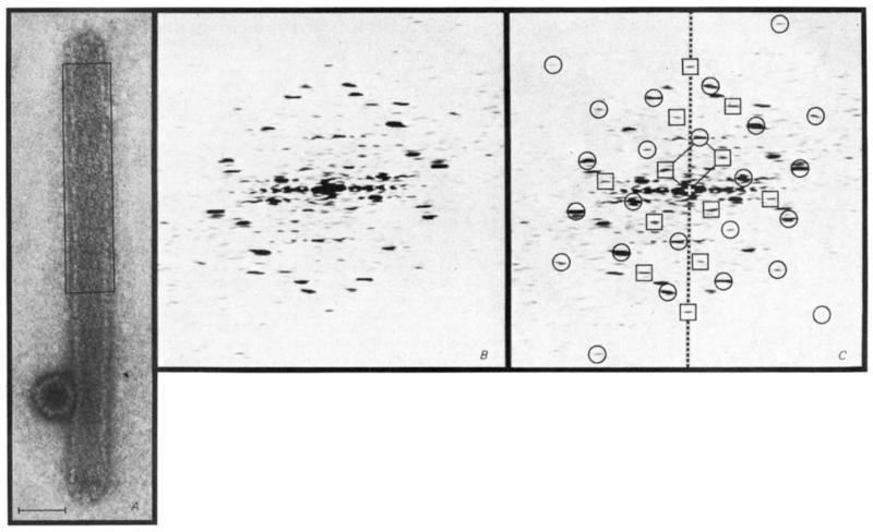
Fig. 1.
Indexing of the diffraction pattern from a polyoma virus hexamer tube image shows that the true unit cell is twice as large as the simple hexagonal cell previously described4 The electron micrograph (A) is representative of the most frequent class of hexamer tube. The tube, capped at both ends, appears uniformly flattened except where it is supported by the adjacent icosahedral capsid. Superposition of the front and back lattices gives rise to a characteristic criss-cross pattern of near axial rows of capsomeres. Scale bar, 500 Å. The diffraction pattern computed from the area boxed in A is shown in B and again in C with indexing of the spots arising from the top layer (away from the grid) of the flattened tube. Circles indicate spots from the simple hexagonal unit cell, which would contain only one capsomere, and the squares mark additional spots from the repeating unit of double this size. Spots from the bottom side (not marked) lie on a lattice related to that from the top by approximate mirror symmetry about the meridional axis (dashed line). The reciprocal lattice unit cell for the top layer is marked at the centre of the pattern.

