Fig. 2.
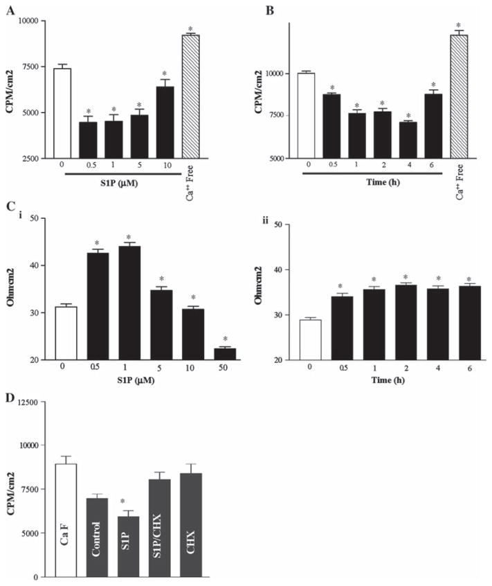
Epithelial cell barrier function after treatment with S1P. A Paracellular permeability. Dose response of cells exposed to S1P for 4 h. B Time course response to S1P (0.5 μM). (C) TEER data for conditions replicating A (i) and B (ii). Values are means ± SE of data from six samples. * P <0.05 compared with control IECs unexposed to S1P. D Paracellular permeability comparing S1P (0.5 μM) with co-incubation of cycloheximide (CHX—25 μg/ml) alone and CHX + S1P. * P <0.05 compared with control IECs unexposed to S1P. The results shown are means from six experiments. All cells were viable (early passage) at the time of experimentation

