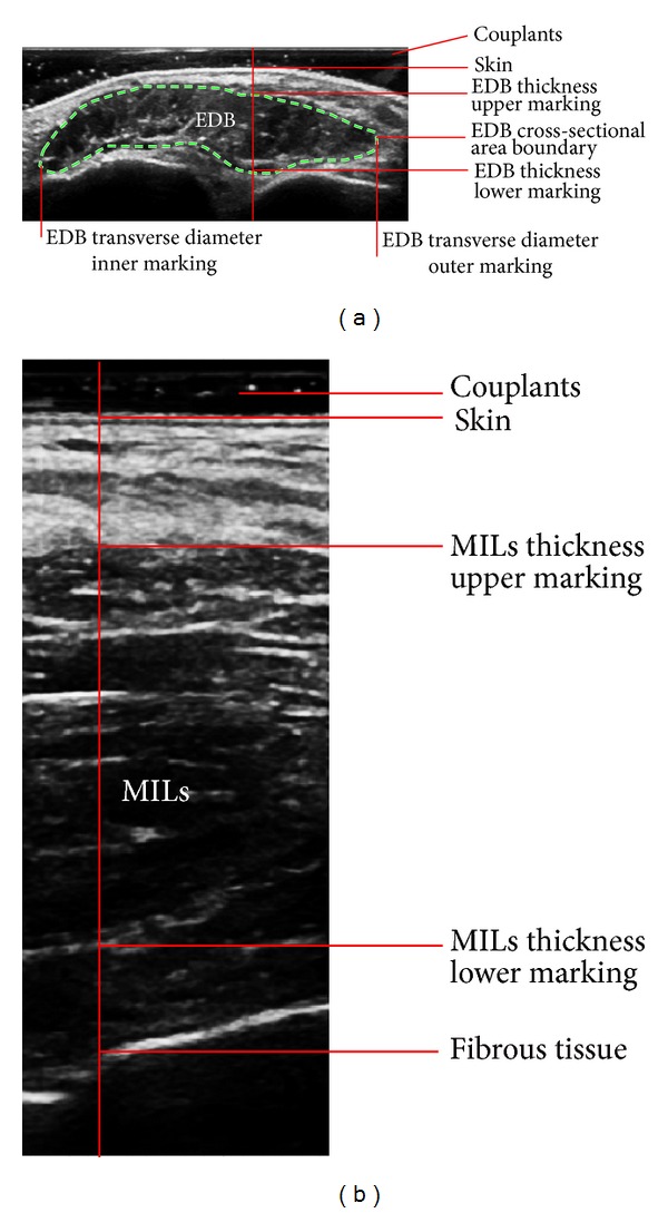
Figure 1.

Ultrasonographic images of the extensor digitorum brevis muscle (EDB) and the muscles of the first interstitium (MILs). (a) A representative ultrasonic image of EDB, along with the enthesis (indicating lines with annotations) and boundary (dashed line) of the measured transverse diameter, thickness, and cross-sectional area. (b) A representative ultrasonic image of MILs and the enthesis (indicating lines with annotations) of the measured thickness.
