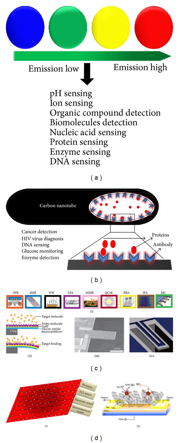
Figure 2.

Schematics for some recent advancement in biosensors applicable in tissue engineering. (a) Variation of color in quantum dots (blue, green, yellow, and red) based on their emission wavelength. (b) Carbon nanotube based biosensor for detecting various cell secreted biomolecules from tiny amount of sample. (c) Some MEMS based biosensors: (i) SPR: surface-plasmon resonance; SMR: suspended microchannel resonator; NW: nanowire; LFA: lateral flow assay; MRR: microring resonator; QCM: quartz crystal microbalance; BBA: biobarcode amplification assay; IFA: immunofluorescent assay; MC: microcantilever. (ii) static-mode surface-stress sensing by a MEMS device (iii) scanning electron micrograph of dynamic mode MEMS device and (iv) suspended microchannel resonator (SMR). (d) (i) Graphene and its derivatives (graphene oxide, graphene quantum dots) based sensors. (ii) Vertically-oriented graphene based field effect transistor-sensor by direct growth of VG between the drain and the source electrodes. (c) and (d) (ii) reproduced from [13] and [132], respectively, with permission from Nature Publishing Group.
