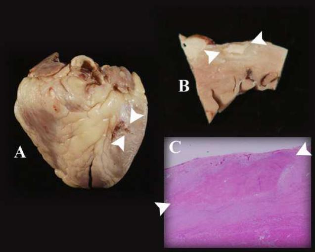
Figure 4.
Gross, sectioned, and microscopic examination of the heart displays the epicardial ablation lesions. In each panel, the ablation lesions are indicated by the two white arrowheads. A: The lesions are seen at the basal anterolateral aspect of the whole heart. The lesions to the left of the arrowheads were superficial, as demonstrated in panel B. B: Section through the area ablation, revealing dense scar of ablation surrounded by scar from sarcoidosis. The depth of the lesion is 2 mm. C: The microscopic cross sectional examination shows that the ablation lesion is in an area affected by sarcoidosis. There is normal myocardium at the lower border of the panel.

