
Figure 1.
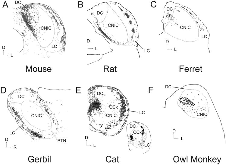
Illustration of the pattern of CC input to the IC after an injection of an anterograde tracer into the primary auditory cortex of six different species. All sections are coronal except D) which is sagittal. Sections redrawn from the following publications: A) Mouse (Torii et al. 2012), B) Rat (Saldaña, Feliciano and Mugnaini 1996), C) Ferret (Bajo et al. 2007), D) Gerbil (Budinger et al. 2013), E) Cat (Winer et al. 1998) and F) Owl Monkey (Fitzpatrick and Imig 1978). Inset in E is a caudal section chosen to illustrate the patchiness of the CC system taken at a point 21% rostral to the caudal pole (100%=anterior-most portion of IC). CCx = caudal cortex of the IC. PTN = paracentral tectal nuclei.