Fig. 1.
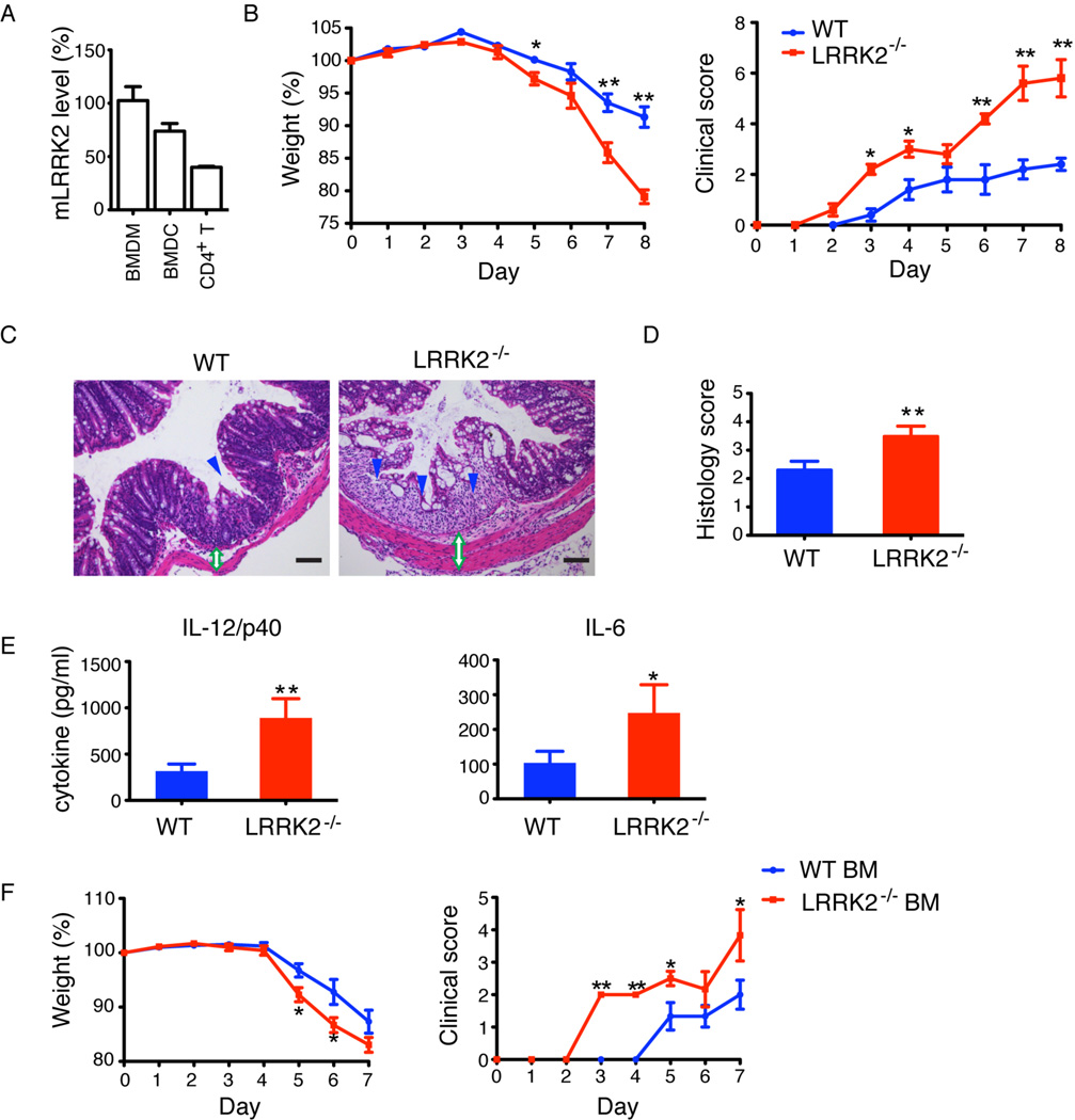
LRRK2 deficiency exacerbates experimental colitis in mice. (A) Relative levels of LRRK2 mRNA measured by qRT-PCR in bone-marrow derived macrophages (BMDM)(set as 100%), bone-marrow derived dendritic cells (BMDC) and CD4+ T cells. β-actin was used for total mRNA normalization. (B) Mean body weight as a percent of starting weight (left panel) and mean clinical scores (right panel) of WT (blue, n=5) and LRRK2−/− (red, n=5) mice treated with 3% DSS. *P < 0.05. **P < 0.02. (C) Hematoxylin and eosin (H&E) staining of colonic sections. Loss of epithelial crypts (blue arrowhead), transmural inflammation and thickened intestinal wall (green open double arrow) are noted. Scale bar, 200 µm. (D) Mean of histological scores of colon sections. **P < 0.02. (E) Serum cytokines at Day 8. *P < 0.05, **P < 0.02. (F) Mean body weight (left panel) and mean clinical scores (right panel) of WT mice that received either WT or LRRK2−/− bone marrow (BM) (n=6). Data are representative of three independent experiments (A–E) and two independent experiments (F). Error bars represent standard errors of the mean (SEM).

