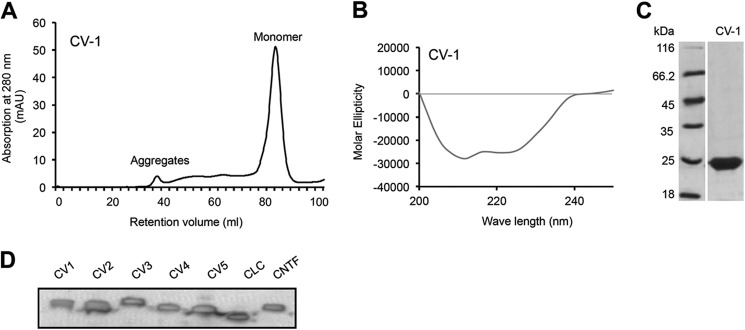
FIGURE 2.

Pure CV-1 is monomeric and correctly folded. A, CV-1 was expressed in E. coli, purified via nickel-nitrilotriacetic acid agarose, and refolded. Finally, CV-1 was purified and analyzed by size-exclusion chromatography. CV-1 was eluted as a monomer. B, CD revealed the correct folding of CV-1. C, Coomassie stains of CV-1. CV-1 was separated by SDS-PAGE and visualized by Coomassie stain. D, Western blot analysis of purified CV-1 to CV5, CLC, and CNTF. Recombinant proteins were separated by SDS-PAGE and detected by Myc-mAb.
