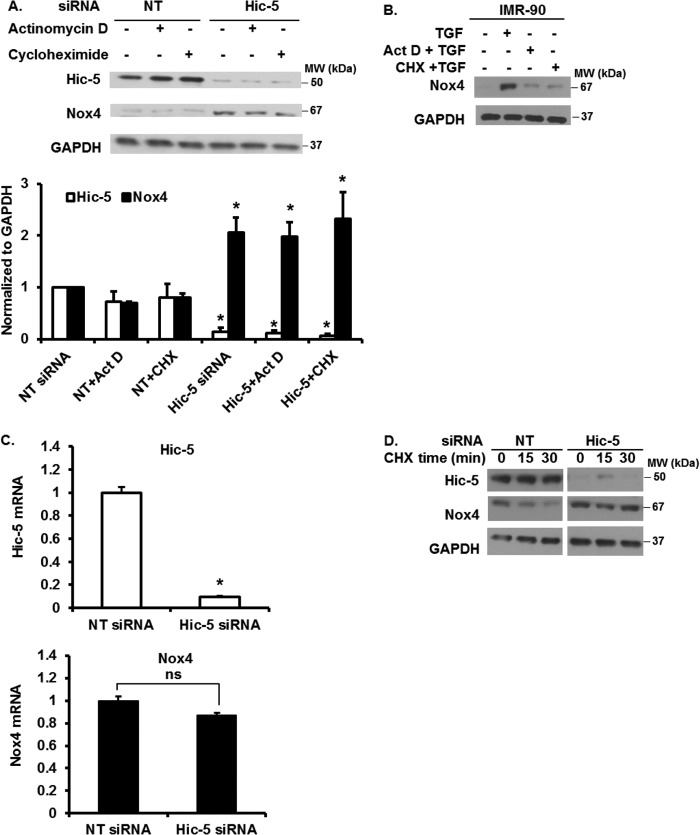
FIGURE 3.
Transcription and translational mechanisms do not regulate Nox4 expression. A, IMR-90 cells silenced in Hic-5 or control (NT siRNA) cells were serum-starved and treated without or with 0.05 μg/ml of actinomycin D or 1 μg/ml of cycloheximide for 2 h. Cells were lysed, and the levels of Hic-5, Nox4, and GAPDH were determined by Western blotting. Top panel, representative Western blot data. Bottom panel, densitometry analysis results of the Western blot data from three independent experiments. *, p < 0.05, significantly different from the corresponding control (NT siRNA) (n = 3). Data are mean ± S.E. MW, molecular weight. B, IMR-90 cells were serum-starved and treated without or with TGF-β1 and in the presence or absence of actinomycin D (Act D) or cycloheximide (CHX) for 24 h. Cells were lysed, and the levels of Nox4 and GAPDH were determined by Western blotting. C, Hic-5 and Nox4 mRNA levels in IMR-90 cells silenced in Hic-5 or control (NT siRNA) cells were determined by real-time PCR (n = 3). Data are mean ± S.E. *, p < 0.005, significantly different from the control (NT siRNA); ns, non-significant. D, IMR-90 cells silenced in Hic-5 or control (NT siRNA) cells were serum-starved and treated with cycloheximide for the indicated times. Cells were lysed, and the levels of Hic-5, Nox4, and GAPDH were determined by Western blotting.

