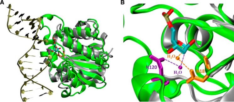
FIGURE 11.

Comparison of interactions between Tth UDGb-N120 with water and E. coli MUG-N18 with water. A, superimposition of UDGb-AP structure (Protein Data Bank code 2DEM; green) with E. coli MUG structure (Protein Data Bank code 1MUG; silver). The AP site is colored by atom type. The two structures were superimposed using the program VMD. B, close-up view of Tth UDGb-N120-water and E. coli MUG-N18-water interactions using the same coloring scheme described in A. Asn120 and the interacting water in Tth UDGb structure are shown in purple. Asn18 and the interacting water in the MUG structure are shown in orange.
