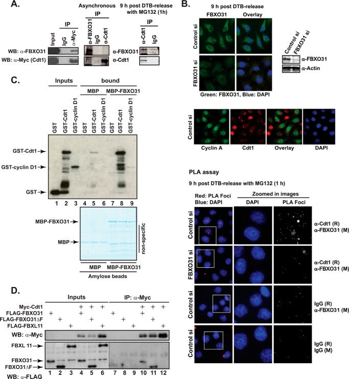
FIGURE 1.
FBXO31 interacts with Cdt1. A, HEK293T cells were transfected with Myc-Cdt1 expression construct. Cell lysates were immunoprecipitated (IP) with either mouse IgG control or anti-Myc antibody. Input and IP samples were Western-blotted (WB) with anti-Myc or anti-FBXO31 antibodies (left panel). For endogenous immunoprecipitation, either asynchronous HeLa cells or cells synchronized with a double-thymidine block and harvested 9 h after release were used. The synchronized cells were treated with MG132 for 1 h before harvest. Total cell extract was subjected to immunoprecipitation using anti-Cdt1, anti-FBXO31 antibodies, or rabbit IgG. The immunoprecipitates were analyzed by Western blotting with anti-FBXO31 and anti-Cdt1 antibodies (right panel). B, HeLa cells were transiently transfected with control or FBXO31 siRNA (siRNA-1), synchronized with a double-thymidine block, and fixed onto coverslips 9 h after release. Samples were treated with MG132 for 1 h before fixation where indicated on the figure. Cells were probed with anti-FBXO31, anti-Cdt1, and anti-cyclin A antibodies or mouse (M) or rabbit (R) IgG alone or in combination as indicated on the figure. Antibodies were either detected by fluorescently labeled secondary antibodies or a proximity ligation assay kit as marked. C, GST, GST-Cdt1, or GST-cyclin D1 proteins expressed and purified from bacteria were incubated with bacterially-expressed MBP or MBP-FBXO31 fusion proteins attached to amylose resin. The complexes were washed and eluted in 1× protein-loading buffer and either analyzed by Western blotting with anti-GST antibody (upper image) or resolved by SDS-PAGE and visualized by colloidal Coomassie staining (lower image). Note a low level of nonspecific GST-Cdt1 binding to MBP-beads in lane 5. D, HEK293T cell lysates expressing FLAG-FBXO31, FLAG-FBXO31ΔF, or FLAG-FBXL11 either alone or together with Myc-Cdt1 were immunoprecipitated with anti-Myc antibody. Inputs and IP samples were Western-blotted with anti-Myc or anti-FLAG antibodies. Note different exposures of the same blot with input and IP samples probed with anti-FLAG antibody were used for clarity.

