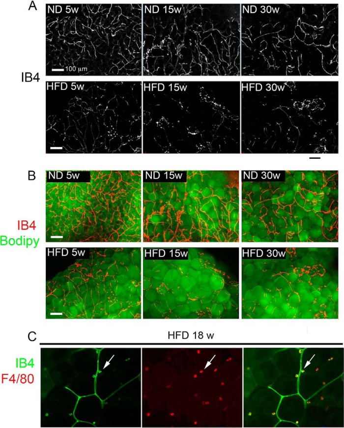
FIGURE 5.
Effect of HFD on adipose tissue capillary network integrity. A, maximal intensity projections of 25 image stacks acquired at 10-μm intervals (250-μm thickness) of IB4-stained fragments of adipose tissue from mice fed ND or HFD (top or bottom panels, respectively) for 5, 15-, or 30 weeks (left, middle, and right panels, respectively). B, superimposition of images in A over corresponding Bodipy-stained images. C, optical section through a fragment of adipose tissue from 18-week HFD-fed mouse double-stained with IB4 (left panel) and antibody to F4/80 (middle panel). Right panel is the overlap of IB4 and F4/80. Arrows point to examples of a single cells positive for both IB4 and F4/80.

