FIGURE 7.
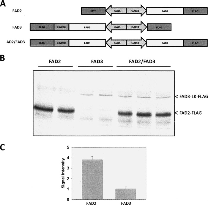
Protein quantification of FAD2 and FAD3 in yeast cells. A, schematic representation of the plasmid constructs made for expression of genes in the yeast cells. A 36-amino acid flexible peptide linker (GGSAGGSGSGSSGGSSGASGTGTAGGTGSGSGTGSG) was inserted between the C terminus of FAD3 and the FLAG epitope tag. B, yeast cells from strain YPH499 expressing Fad2 or Fad3 or coexpressing Fad2 and Fad3 (Fad2/Fad3) in pESC-Ura vectors were grown in medium containing 0.1 mm oleic acids and 0.1 mm linoleic acids at 30 °C for 4 h followed by incubation at 18 °C for 24 h. Extracts were separated by SDS-PAGE, immunoblotted, and visualized with the use of anti-FLAG antibodies. C, quantitation of Western blotting signal for FAD2-FLAG and FAD3-LK-FLAG.

