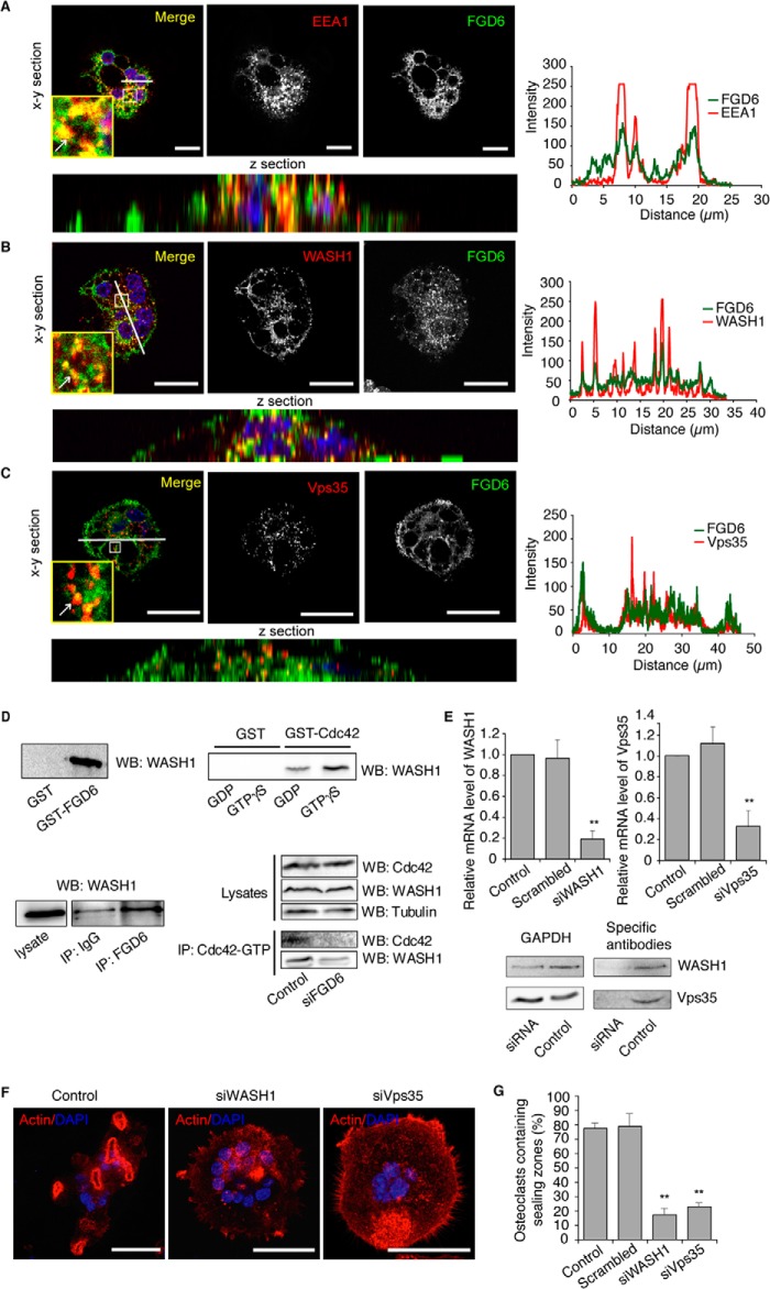
FIGURE 5.
FGD6 interacts with WASH1 on endosomes. Osteoclasts expressing GFP-FGD6 (green) were grown on ODs and stained with DAPI (blue) and anti-EEA1 (A), anti-WASH1 (B), or anti-Vps35 (C) (red), and analyzed by confocal microscopy. Bars, 20 μm. Arrows show early endosomes around transcytotic vesicles. The corresponding fluorescence intensity profiles along the white line are shown. D, osteoclast lysates were incubated with GST, GST-FGD6(1–1039), or GST-Cdc42 loaded with GDP or GTPγS, or with anti-FGD6 antibodies. After pull down, the interacting material was analyzed by Western blotting (WB) using anti-WASH1. GTP-Cdc42 was immunoprecipitated from lysates of osteoclasts depleted or not from FGD6, and after Western blotting the immunoprecipitates (IP) were probed for Cdc42 and WASH1. E–G, osteoclasts grown on ODs were mock-transfected, or transfected with scrambled siRNAs or with siRNAs targeting WASH1 or Vps35. E, the expression levels were determined by quantitative RT-PCR. Osteoclasts were also collected and lysed. Lysates were analyzed by SDS-PAGE followed by Western blotting using antibodies directed against the indicated gene products. F, osteoclasts were also stained with phalloidin (red) and DAPI (blue) and analyzed by confocal microscopy. Bars, 50 μm. G, the number of osteoclasts with sealing zones was quantified. Values are mean ± S.D. from 3 experiments (n = 200 osteoclasts per experiment). All groups were compared with control by applying a Dunnett one-way ANOVA test. *, p < 0.05. **, p < 0.01. Representative images are shown. See also supplemental Videos S9 and S10.

