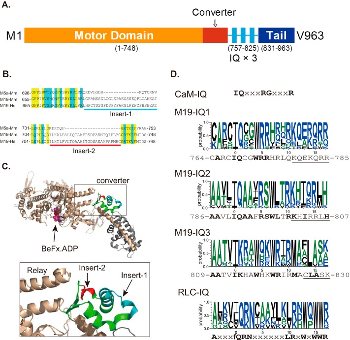
FIGURE 1.
Diagram of Myo19 structure and sequence alignment of Myo19. A, predicted structure of Myo19. The diagram was not drawn to scale. B, sequence alignment of the converter of myosin-5a and Myo19 reveals two inserts in the converter of Myo19. Yellow and blue shades indicate the identical and conserved residues, respectively. Myo5a-Mm, mouse myosin-5a (GI: 115511052); M19-Mm, mouse Myo19 (GI: 254939539); M19-Hs, human Myo19 (GI: 254939537). C, location of the two Myo19-specific inserts in the crystal structure of myosin-5a (PDB code 1W7J). Upper, the head domain of myosin-5a; lower, the expanded view of the converter. The converter is colored green with insert 1 in cyan and insert 2 in red. Insert 1 and 2 are adjacent to each other and insert 2 is adjacent to the tip of the relay helix. D, consensus sequences of CaM-binding IQ motif, IQ motifs of Myo19, and RLC-binding IQ motif of myosin-2 and -18. CaM-IQ, consensus sequence of the six IQ motifs of mouse myosin-5a (GI: 115511052). M19-IQ1, -IQ2, and -IQ3, sequence logo of the consensus sequence of the three IQ motifs of 11 class XIX myosins, including mouse (GI: 254939539), rat (GI: 254939541), human (GI: 254939537), cattle (GI: 254939551), elephant (GI: 344285323), rabbit (GI: 291405654), cat (GI: 410980571), whale (GI: 466018728), chicken (GI: 513216545), Xenopus laevis (GI: 82178330), and zebrafish (GI: 189519181). The sequence shown at the bottom is mouse Myo19. Conserved residues are shown in bold. Underlined residues are not essential for RLC binding (for details see ”Discussion“). RLC-IQ, sequence logo of the consensus sequence of IQ2 motifs of five muscle myosin-2 and two myosin-18, including scallop muscle myosin (GI: 5611), human non-muscle myosin-2a (GI: 47678583), -2b (GI: 219841954), -2c (GI: 116284394), smooth muscle myosin (GI: 46486992), myosin-18a (GI: 24660442), and myosin-18b (GI: 219841774). Sequence logos were generated by an online program, WebLOGO.

