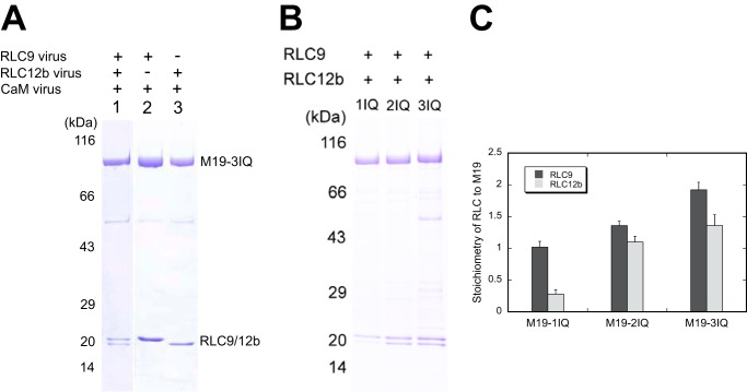
FIGURE 4.
Identification of the light chain binding sites in Myo19. Myo19-truncated constructs were co-expressed with different groups of light chains. The purified Myo19 samples were subjected to SDS-PAGE (4–20%) and Coomassie Blue staining. A, SDS-PAGE of purified M19-3IQ coexpressed with the different light chains (indicated at the top of the gel) in Sf9 cells. B, SDS-PAGE of purified M19-1IQ, -2IQ, and -3IQ coexpressed with RLC9 and RLC12b. C, stoichiometry of RLC9 and RLC12b to Myo19 heavy chain. Quantifications of the light chain and heavy chain were done with ImageJ software. The molar ratio of RLC versus the heavy chain was calculated based on the corresponding molecular weight masses. Values are mean ± S.D. from three independent assays.

