FIGURE 8.
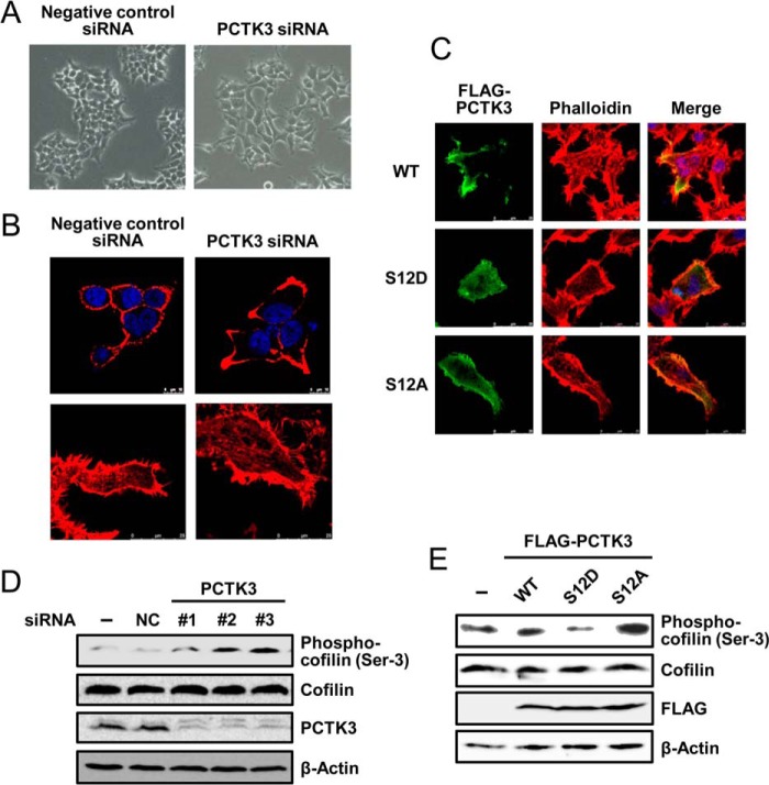
Suppression of PCTK3 induces actin cytoskeletal changes through cofilin phosphorylation. A and B, HEK293T cells were transfected with negative control siRNA or PCTK3 siRNA. After 48 h, cells were fixed and incubated with Alexa 555-conjugated phalloidin (B, red). Hoechst nuclear staining is represented in blue. C, HEK293T cells were transfected with PCTK3 siRNA for 48 h and then transfected with FLAG-tagged mouse PCTK3 wild type (WT) or S12D or S12A mutants. After 24 h, cells were fixed and incubated with mouse anti-FLAG antibody. The primary antibody was visualized with Alexa Fluor 488-conjugated anti-mouse IgG followed by confocal microscopy. Fluorescence for FLAG-PCTK3 (green) and F-actin (Alexa 555-conjugated phalloidin staining, red) are shown with merged images (merge is in yellow). Hoechst nuclear staining is represented in blue. D, cell lysates of PCTK3 knock-down HEK293T cells were subjected to immunoblotting with anti-PCTK3, anti-cofilin, and anti-phospho-cofilin (Ser3) antibodies. Expression of β-actin was used as a loading control. E, HEK293T cells were transfected with FLAG-PCTK3 (WT or S12D or S12A mutant). After 24 h, cell lysates were subjected to immunoblotting using anti-phospho-cofilin (Ser3), anti-cofilin, and anti-FLAG antibodies.

