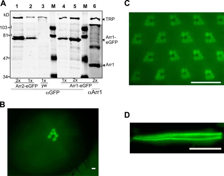
FIGURE 1.
Characterization of transgenic flies expressing Arr1-eGFP in R1–6 photoreceptors. A, Western blot analysis. Shown is a representative Western blot comparing the levels of Arr1-eGFP and Arr2-eGFP as well as the relative levels of Arr1-eGFP and endogenous Arr1 in transgenic flies. Lanes 1 and 2 contain head extracts from Arr2-eGFP expressing flies, whereas lane 3 was from the parental strain yw. Lanes 4–6 contain head extracts from Arr1-eGFP-expressing flies. Double amounts of extracts were loaded in lanes 1, 5, and 6. The estimated size for Arr1-eGFP is about 71 kDa, and that of Arr2-eGFP, about 75 kDa. Arr1 corresponds to a polypeptide of 44 kDa, whereas TRP (transient receptor potential) is a polypeptide of 150 kDa, which was used as a loading control. Protein size markers (M) are indicated on the left. B, enrichment of Arr1-GFP in the rhabdomeres can be detected as fluorescent “deep pseudopupil” in adult eyes. C, subcellular localization of Arr1-eGFP in wild-type photoreceptors under blue light illumination. D, Arr1-eGFP is highly concentrated in rhabdomeres but also present in the cytoplasm of isolated photoreceptors. Scale bar, 20 μm.

