FIGURE 2.
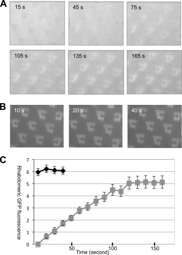
Kinetics of the light-dependent translocation of Arr1-eGFP and Arr2-eGFP in adult retinas. A, subcellular localization of Arr1-eGFP during continued blue light illumination. Selected time-lapse images are shown (see supplemental Movie 1). Arr1-eGFP initially was in the cytosol but began to migrate to rhabdomeres at ∼45 s after the blue light was switched on. Translocation reached a steady state after around 165 s. B, light-dependent redistribution of Arr2-eGFP. The light-dependent translocation of Arr2-eGFP occurred much faster than that of Arr1-eGFP. C, a comparison of translocation kinetics of Arr2-eGFP (black) and Arr1-eGFP (gray) in wild-type background. Data are represented as the mean ± S.E. (n = 3).

