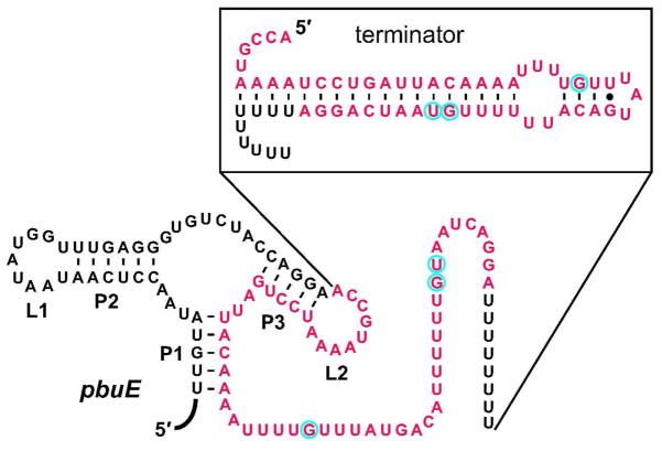
Figure 5.
Sequence and secondary structure of the pbuE adenine riboswitch in B. subtilis. In the absence of adenine, the riboswitch assembles a transcription terminator stem (shown in the box above). Lines from the box to the pbuE secondary structure designate the beginning and end points of the nucleotides shown in the box. The red nucleotides depict the deletion mutation found in one G6-resistant strain. The blue circles each represent one G6-resistant mutant strain in which the corresponding point mutation was found.

