Figure 2.
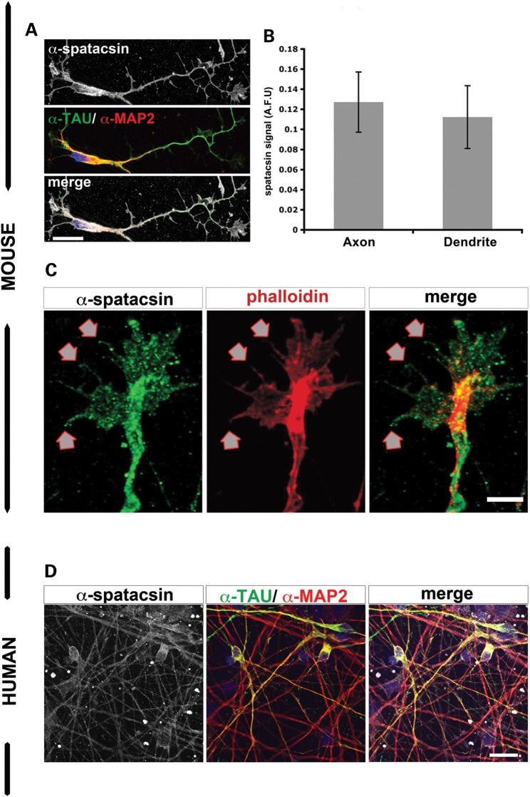
Spatacsin was present in the most distal tips of neurites of human pluripotent stem cell-derived neurons and mouse cortical neurons. (A) Mouse cortical neurons showed spatacsin expression (gray) together with the axonal marker α-TAU (green) and the dendritic marker α-MAP2 (red). Scale bar = 20 µm. (B) Graph for spatacsin expression as arbitrary fluorescent units (AFU) in axons (TAU+ neurites) and dendrites (MAP2+ neurites) of mouse cortical neurons; data represented as mean ± SD (P > 0.05); n ≥ 50 neurons per experimental condition were evaluated. (C) Spatacsin was observed in filopodia and membrane protusions (arrows) of growth cones of mouse cortical neurons. Scale bar = 5 µm. (D) Immunofluorescence analysis of HUES6-dNeuron cultures showed α-spatacsin (gray) overlapped with α-MAP2 (red) and α-TAU (green) markers. Scale bar = 50 µm.

