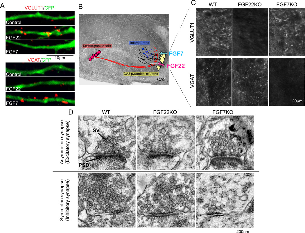
Figure 4. FGF22 and FGF7 promote glutamatergic and GABAergic synaptic differentiation, respectively.
(A) Overexpression of FGF22 and FGF7 selectively promotes VGLUT1 or VGAT clustering on the FGF-expressing hippocampal neurons (labeled with GFP) in culture. (B) CA3 pyramidal neurons, by which FGF22 and FGF7 are expressed, receive excitatory inputs from dentate granule cells (and CA3 and entorhinal cortical neurons) and inhibitory inputs from GABAergic interneurons in CA3. (C) Impaired glutamatergic and GABAergic presynaptic differentiation in CA3 of FGF22KO and FGF7KO mice, respectively. Glutamatergic and GABAergic presynaptic differentiation was assessed by measuring the clustering of VGLUT1 or VGAT. Pictured areas correspond to the boxed area in (B). (D) Representative electron microscopic pictures of asymmetric (excitatory) and symmetric (inhibitory) synapses in CA3 of wild-type (WT), FGF22KO and FGF7KO mice. Relative to WT synapses, excitatory synaptic vesicles are more diffusely distributed in FGF22KO synapses, while less inhibitory synaptic vesicles are observed in FGF7KO synapses. Adapted from Terauchi and others (2010). SV: synaptic vesicle, PSD: postsynaptic density.

