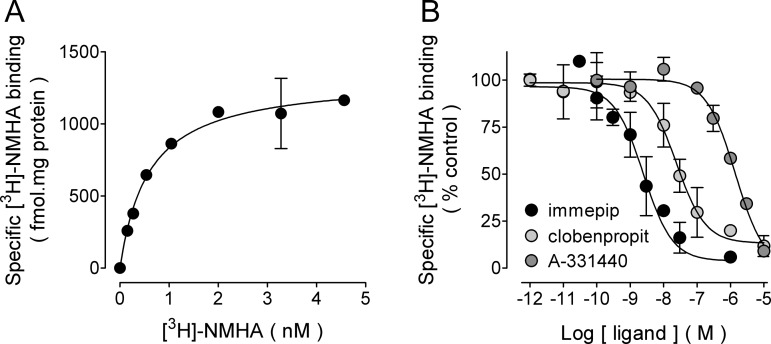
Figure 3.
Binding of N-α-[methyl-3H]-histamine ([3H]-NMHA) to membranes from rat globus pallidus synaptosomes. (A) Saturation binding. Membranes were prepared as described in Methods and then incubated with the indicated concentrations of [3H]-NMHA. Specific receptor binding was determined by subtracting the binding in the presence of 10 μM histamine from total binding. Points are means ± SEM from triplicate determinations from a single experiment, which was repeated a further twice. The line drawn is the best fit to a hyperbola. Best-fit values for the equilibrium dissociation constant (Kd) and maximum binding (Bmax) are given in the text. (B) Inhibition by the H3R agonist immepip and the antagonists/inverse agonists clobenpropit and A-331440. Membranes were incubated with ∼1.5 nM [3H]-NMHA and the indicated drug concentrations. Values are expressed as the percentage of control specific binding and are means ± range from duplicate determinations from a representative experiment. The line drawn is the best fit to a logistic equation for a one-site model. pKi values calculated from the best-fit IC50 estimates are given in the text.

