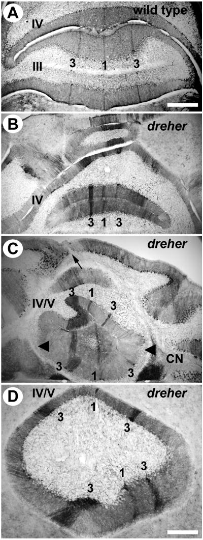
Figure 3.
Zebrin II is expressed in parasagittal stripes in the anterior zone (AZ) vermis of dreher mice. (A) At least three zebrin II immunopositive stripes are reproducible in the AZ of +/+ cerebella. In lobule III, P1+ is located at the midline and is heavily reactive for zebrin II. Two P3+ stripes are located approximately 500 μm lateral to P1+ and are also heavily reactive for zebrin II. P1- is broad and is located directly between P1+ and P3+. In lobule IV, P1- tapers and the distance between P1+ and P3+ are reduced accordingly. (B) In dreher, P1+ is central and is flanked by two P3+ stripes located 400-500 μm laterally. The width of P1- is considerably reduced. (C) In lobule IV, zebrin II immunopositive stripes appear deflected from the midline of the cerebellum. The zebrin II P1+ stripe and the morphological midline seen from lobules at the cerebellar surface (arrow) are not aligned. Despite the twisting of the stripes, which no longer run parasagitally, the distance between them is relatively normal. Irregularities are observed in the width of individual stripes (e.g., the P3+ stripe on the left side is wider at this level of the AZ in this individual and the P1- on the left is concomitantly reduced. (D) Higher magnification view of lobules IV/V, illustrating the twisted architecture of the AZ stripes P1+ and P3+. Roman numerals indicate cerebellar lobules. Scale bar = 500 μm in A (also applies to B and C); 250 μm in D.

