Figure 7.
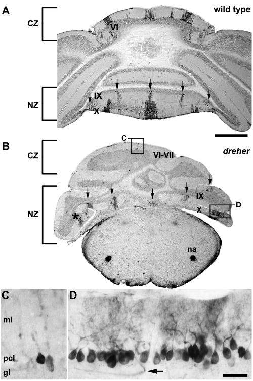
The expression of HSP25 reveals parasagittal stripes in the nodular zone (NZ) of dreher mice. Similar to wild type mice (A), at least five HSP25 immunoreactive stripes are detected in lobules IX and X of dreher mice (B). One stripe is located at the midline and is flanked by two stripes bilaterally (arrows). Note that despite the different rostrocaudal locations of each hemi-vermis, five obvious HSP25 immunoreactive stripes are visible in a single plane. (C) Despite the lateral deflection of lobules, stripes in the caudal CZ are fully differentiated from the stripes in the NZ (higher magnification of the box labeled C in panel B). (D) An example of an individual immunoreactive stripe showing that HSP25 immunoreactive Purkinje cells are organized into a monolayer (higher magnification of the box labeled D in panel B). As in control cerebella, reaction product is heavily deposited in the Purkinje cell somata and dendrites. The arrow points to an immunoreactive blood vessel in the granular layer (for details see Armstrong and Hawkes 2001). Roman numerals indicate cerebellar lobules. Scale bar = 1 mm in A (also applies to B); D = 50 μm (also applies to C).

