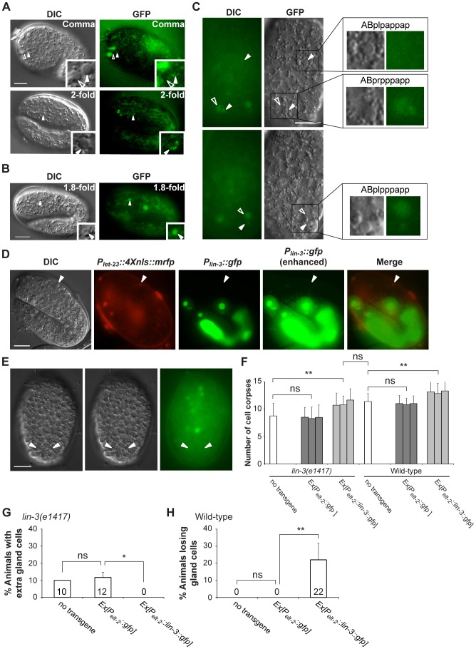
Figure 3. let-23 is expressed in dying cells, whereas lin-3 acts in a cell-nonautonomous manner to promote PCD.
(A and B) Representative DIC and GFP images of embryos carrying (A) the syEx234[Plet-23::let-23::gfp] transgene or (B) the Plet-23::4Xnls::gfp transgene. The white arrowheads indicate cell corpses expressing LET-23::GFP and the hollow arrowheads cell corpses not expressing LET-23::GFP. Scale bar: 10 µm. The insets show a dying cell at a three-fold higher magnification. (C) The DIC and GFP images of an embryo carrying the Plet-23::4Xnls::gfp transgene. Two different focal planes of the same embryo are shown in the upper and lower panels. The white arrowheads indicate ABpl/rpppapp corpses and ABplpappap and the hollow arrowheads sister cells of ABpl/rpppapp. Scale bar: 10 µm. The insets show the indicated cells at a higher magnification. (D) The expression pattern of Plin-3::gfp and Plet-23::4Xnls::mrfp transgenes in a wild-type embryo. The white arrowheads indicate the same cell corpse expressing Plet-23::4Xnls::mrfp. The image stacks of Plin-3::gfp were merged by maximum intensity projection using Image J. The strong GFP expression is from the co-injection marker Pelt-2::gfp. Scale bar: 10 µm. (E) Plin-3::gfp is not expressed in ABpl/rpppapp corpses. The image stacks of Plin-3::gfp were merged by maximum intensity projection using Image J. Scale bar: 10 µm. (F–H) Intestine-specific expression of LIN-3 rescues the cell death defect of the lin-3 mutant and causes ectopic cell deaths in the wild-type. Embryonic cell corpses in the indicated genotypes were scored at the 1.5-fold stage (n≥30), and the numbers of gland cells were scored at the L4 stage (n≥40). Three independent stable transgenic lines were analyzed. *indicates P<0.05 and **P<0.001 in a two-tailed t-test (F) or fisher's exact test (G and H). ns indicates no statistical difference (p>0.05).

