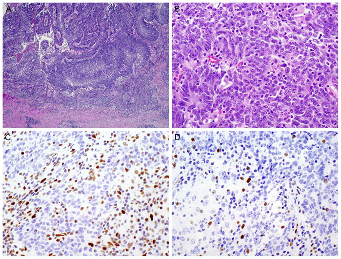
FIGURE 2.
Patient 2. (A) Low-power appearance of an undifferentiated, medullary-type carcinoma with vague papillary fronds projecting into the appendiceal lumen. (B) Sheet-like growth at high-power. (C) Loss of MSH2 expression in the tumor, but intact expression in the tumor-infiltrating lymphocytes. (D) Loss of MSH6.

