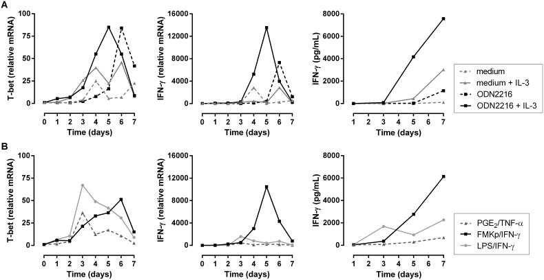
Figure 7. Differential Th1 polarizing capacity of differently matured plasmacytoid DC.
pDC, isolated from fresh blood, were stimulated overnight with different cocktails and 24 h later autologous naive CD4+ T cells were added and the expression of T-bet and IFN-γ and secretion of IFN-γ were monitored during 7 days. (A) pDC were stimulated overnight with IL-3 (full gray line), ODN2216 (dashed black line) or with a combination of both (full black line). (B) Comparison of the capacity of differently matured pDC to induce Th1 polarization. pDC were incubated with PGE2/TNF-α (dark gray triangle), LPS/IFN-γ (light gray circle) or FMKp/IFN-γ (black square) cocktail in the presence of IL-3. Representative data from 2 independent experiments are shown.

