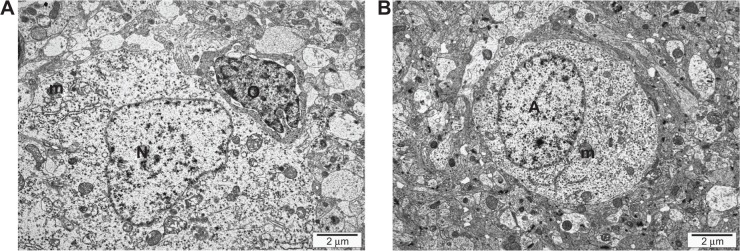
Figure 3.

Transmission electron microscope image of the ultrastructure of thin sections of cerebrum samples of a 20-day-old chicken embryo from the control group. (A) Mitochondrion (m), nuclei of nerve cells (N), nuclei of oligodendroglia (O). (B) Nuclei of astrocytes (A), mitochondrion (m).
