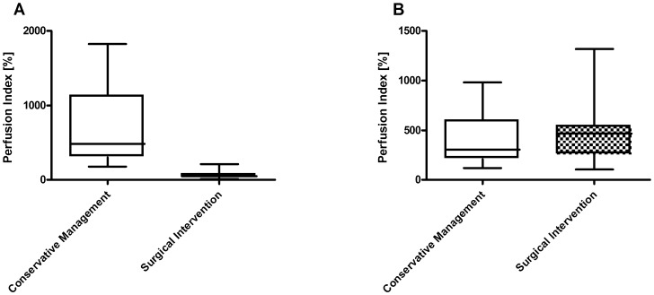
Figure 1. Perfusion at central area (A) and peripheral area (B) of the extravasation lesion.
Box plots represent cutaneous blood flow as assessed by the perfusion index in patients with conservative clinical management (N = 22) versus those requiring subsequently surgical intervention (N = 7); *statistical significance p<0.05.

