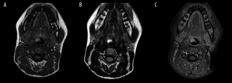
Figure 1.
Axial T1-weighted (A) and T2-weighted (B) images show a lobulated mass along the free edge of the epiglottis to the right of midline. The mass was hyperintense. No intrinsic signal abnormality of the adjacent vallecula or tongue base was noted. Post contrast administration, the mass showed no enhancement (C).

