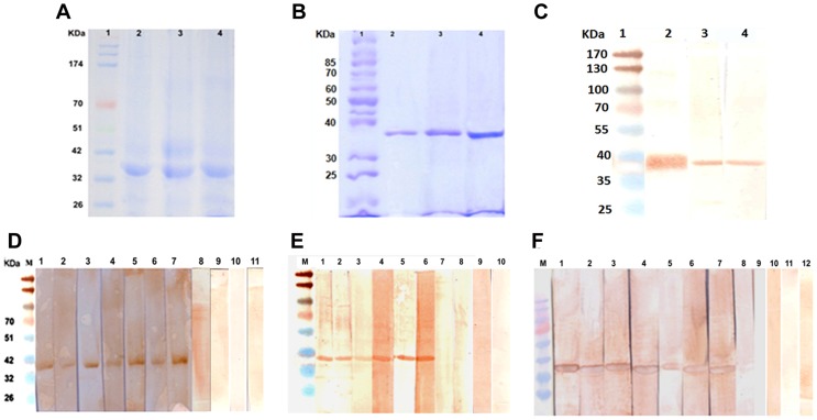
Figure 1. Cloning and expression of cpa, cpb and cpc.
A, Coomassie blue stained protein fractions showing overexpression of cpa, cpb and cpc in E. coli BL21 (DE3). Lane 1, prestained molecular weight marker; lane 2, cpa; lane 3, cpb; lane 4, cpc. B, Coomassie blue staining of 10% SDS-PAGE showing purified proteins. Lane 1, molecular weight marker; lane 2, rCPA; lane 3, rCPB and lane 4, rCPC. C, recombinant proteins probed with anti-polyhistidine antibody after Western blot. Lane1, molecular weight marker; lane 2, rCPA; lane 3, rCPB and lane 4, rCPC. D–F, Plasma recognition of L. donovani CP. Immunoblot analysis of recognition of recombinant CPA (D), CPB (E) and CPC (F) by human blood plasma from infected (odd numbered lanes 1–7) and cured (even numbered lanes 1–7) individuals suffering from VL. Lane 8–12, endemic controls. M, molecular weight marker.

