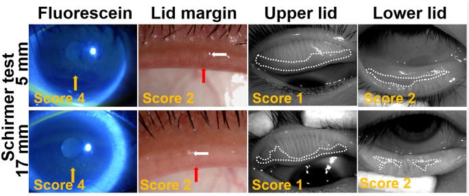
Figure 3. Representative images of fluorescein staining, lid margin and meibography in patients with or without tear volume deficiency in long time VDT group.
The score of each image is noted at bottom left. The upper panel shows eyes from low tear volume subgroup: the right eye from a 29-year-old male, VDT work time: 12 h/d; OSDI: 54.55; BUT: 7 s; Schirmer test: 5 mm; the meibum score was 2. The bottom panel shows eyes from normal tear volume subgroup: the right eye from a 37-year-old male, VDT work time: 6 h/d; OSDI: 40.00; BUT: 5 s; Schirmer test: 17 mm; the meibum score was 1. Corneal fluorescein staining is noted by yellow arrows. Vascular engorgement is outlined by red arrow and plugged meibomian gland orifice is outlined by white arrow. The areas of meibomian gland dropout are encircled with dotted white lines.

