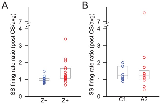
Figure 8. Stronger increase in SS activity after CS in Z+ than Z− bands.

The difference is consistent for both histologically (A) and physiologically (B) located PCs. Each circle represents the median ratio of the SS firing rate for the 100-ms period after a CS to the overall SS firing rate for the individual PCs in the Z− bands or the C1 zone (blue) and in the Z+ bands or the A2 zone (red). The black lines indicate the overall median across each group, and the grey boxes indicate the range from 25% to 75% of each distribution.
